The department of Radiodiagnosis is one of the key departments of the hospital providing basic and advanced radiological diagnostic facilities, as well as therapeutic procedures and interventional radiology. The department follows a dedicated and patient-centric approach for providing timely diagnostic imaging facilities to both inpatients and outpatients being referred from all other departments of the hospital. Basic imaging facilities are provided by the X-ray department, with advanced and state of the art MRI and CT facilities also available 24/7.
The Radiology team comprises of eminently qualified senior professors, associates and assistant professors who are members of well-known radiological societies. They are supported by qualified radiology technicians.
The department has dedicated X-ray facilities, contrast imaging (Barium studies, urography, hysterosalpingography etc.) mammogram facilities, DEXA scans etc. Ultrasound services are available round the clock for a variety of sonological studies, with expert guidance. Basic, as well as advanced ultrasound imaging studies and Doppler studies, are also carried out. Computed Tomography services are available, with state-of-the-art CT machinery. MRI setup provides top-class magnetic resonance imaging services, with cutting edge technology and opinion by expert professors. All sequences with high-resolution thin films are taken for clarity and confidence in reporting. Contrast CT and MRI studies are carried out to augment the accuracy and scope of the diagnosis.
Radiology department has a dedicated PACS system in place which provides a permanent database for all the imaging studies and helps in record maintenance and patient follow up.
The department, with a focus on patient welfare, provides interventional radiological services as well, for performing skilled therapeutic procedures under ultrasonic or CT assisted guidance, which would be otherwise difficult or risky when performed by conventional techniques.
A wide array of ultrasound-guided procedures, such as aspiration of intracavitary fluid collections, abscesses, placement of catheters, focused biopsy of suspicious lesions for maximum accuracy are carried out under expert guidance with utmost attention to patient safety, sterility and relieving patient suffering.
Bedside Xray and ultrasound facilities are provided for immobile or debilitated patients, or those otherwise unable to visit the department, especially those undergoing intensive care or recovering in post-operative wards.

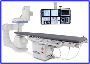
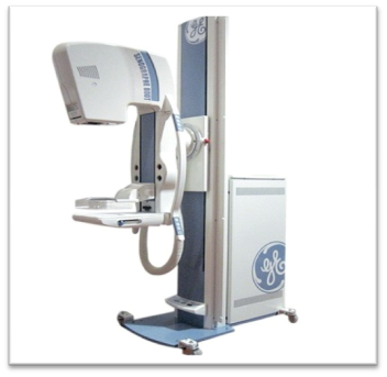
Fluoroscope.
CT Scan.
Mammography.
Registration for Ultrasound Scan.
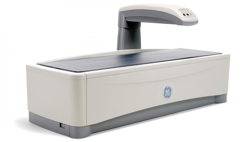
Bone Densitometer.
Fixed X-Ray.
Fixed X-Ray.
C Arm.
Mammography.
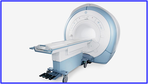
Magnetic resonance imaging is a technique for creating detailed images of the human body, especially of the soft tissues.
In this, a powerful magnetic field is used to align the nuclei of atoms inside the body towards a particular direction, and a variable magnetic field is used to make the atoms resonate, a phenomenon called nuclear magnetic resonance. Then the nuclei of the body's atoms each produce a small magnetic field (depending on the type of tissue). A scanner detects these and creates a computed image. Thus an MRI scan provides very detailed view of the soft tissues of the body, and can even detect diseases in very early stages that other modalities can sometimes fail to pick up.
Diffusion MRI
Diffusion is defined as the movement of molecules from a region of high concentration to a lower one. This technique exhibits images of diseases like stroke and other diseases.
Magnetic Resonance Spectroscopy
This technique operates by detecting changes in metabolites produced in response to certain diseases. Since metabolites produced by abnormal tissues differ from those produced by normal cells, this technique gives additional information

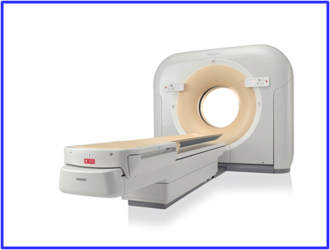
CT or computed tomography is a technique which takes multiple Xray images of the human body, puts them together by means of a powerful computer to create a full cross-sectional view of the internal structures and bones, that is much more powerful than what x-rays alone can tell. It provides valuable diagnostic information in various structural problems and also for many diseases like stroke. The CT scanner takes multiple views of the patient from different directions. All of these are then converted into a composite series of images. Three dimensional images can also be created to provide even more clarity.
CONTRAST CT
Contrast CT is an even more sensitive modality involving the administration of an intravenous, oral contrast (sometimes contrast enema) to provide even more detail and clarity than plain CT alone.

An ultrasound scan uses high-frequency sound waves to create images of the inside of the body. Ultrasound scans, or sonography, are safe because they use sound waves or echoes to make an image, instead of radiation. They are used for diagnosis or treatment. There are different subtypes of ultrasound such as 3D ultrasound and Doppler Ultrasound.
Ultrasound machines transmit high frequency sound waves through the patient’s body using the transducer. These waves may pass or get reflected depending on the density and special properties of each type of tissue. Pathologies can show up when viewing the different organs.
Obstetrics and Gynecology
Ultrasound is very safe and is the most popular obstetric imaging tool, used to view the foetus, detect foetal and maternal diseases and to monitor pregnancies. They are also useful for a variety of gynecological disorders.
Nephrology
Ultrasound is invaluable in imaging the kidneys, detecting problems such as renal stones, their size, and demonstrating any other structural abnormality of the kidneys, ureters and bladder.

Mammograms detect pathologies of the breast, and are done frequently due to the high incidence of breast cancer in middle aged and older women. They are also used to diagnose other breast lesions.
Screening mammogram
This provides a cursory view of the breast and is valuable for wide scale screening of middle-aged women.
Diagnostic mammography
This uses additional radiation to create a detailed image, and is often accompanied by breast ultrasound. .

DEXA stands for dual energy X-ray absorptiometry. A DEXA scan is a high-precision type of X-ray that measures bone mineral density and bone loss. If bone density is lower than normal for age, it indicates a risk for osteoporosis and bone fractures.
The DEXA machine sends a thin, invisible beam of low-dose x-rays with two distinct energy peaks through the bones being examined. One peak is absorbed mainly by soft tissue and the other by bone. The soft tissue amount can be subtracted from the total and what remains is a patient's bone mineral density.DEXA machines feature special software that compute and display the bone density measurements on a computer monitor.
The radiation exposure is low when compared to other tests like CT scan. The entire skeleton is imaged and provides accurate density values. DEXA is also cheaper than many other modalities for bone density estimation.

X-rays are a type of radiation called electromagnetic waves. X-ray imaging shows the parts of the body in different shades of black and white. This is because different tissues absorb different amounts of radiation. Denser tissues show up as whiter regions on the film and less dense tissues such as lungs show up as relatively darker regions. X-rays expose the patient to a small amount of radiation.
Cardio-pulmonary investigation
X-rays provide a quick, cheap methodology for screening of the lungs and detecting heart enlargement or other major problems. Xrays are a valuable screening tool with widespread application